Correctly Label the Following Muscles of the Neck.
An overview Anterior neck muscles. Splenius capitis and splenius cervicis are a pair of superficial muscles in the back of the neck.

Solved Correctly Label The Following Muscles Of The Neck Chegg Com
4 - the skull.

. Under the platysma are two sternocleidomastoid muscles. The muscles of the neck are present in four main groups. Learn the anatomy of a typical human cell.
The muscles of the neck can be divided into two large groups. Test your knowledge of the bones of the full skeleton. Muscles of facial expression include frontalis orbicularis oris laris oculi buccinator and.
The trapezius originates from the skull and spine of the upper back and neck. Label the bony structures of the shoulder and upper limb. 6 - the heart.
All the muscles of the neck are long and thin muscles that act in synergistic agonizing and antagonistic groups to achieve the wide range of movements of the head. Although anchored in the neck their primary functions are to move the shoulder blades and support the arms. Rectus capitis posterior major and Rectus capitis posterior minor attach the inferior nuchal line of the occiput to the C2 and C1 vertebrae respectively.
Lateral neck vertebral muscles. Correctly label the anterior thigh muscles. The neck muscles include the scalenes which attach the cervical vertebrae to the thoracic cage and the sternocleidomastoid which attaches the skull to the thoracic cage.
Below are three with a larger impact. These muscles move the head and neck. Muscles of the Head and Neck.
These are what you feel get tired when you sit close to the screen at a movie theater. Because the muscles are used to show surprise disgust anger fear and other emotions they are an important means of nonverbal communication. Lymphatic drainage of the cervical viscera anterior view.
The neck muscles including the sternocleidomastoid and the trapezius are responsible for the gross motor movement in the muscular system of the head and neck. They insert on the mastoid process of the temporal bone. These muscles give the sides of the neck their shape.
Muscles of the neck. They move the head in every direction pulling the skull and jaw. What is the name of muscle A.
This is a back view of the right leg. Is a paired muscle in the superficial layers of the front part of the neck. It is also an accessory muscle of breathing out and raises the sternum.
The epicranius muscle is also very broad and covers most of the top of. Deep lymphatic drainage of the head and neck lateral view. 2 - the brain.
They can flex or extend the head or can rotate the towards the shoulders. 1 - the skeleton. 3 - the cell.
Several other muscles act on the head and neck. Obliquus capitis superior also extends from the occiput to C1 while obliquus capitis inferior originates. Chin and lower lip.
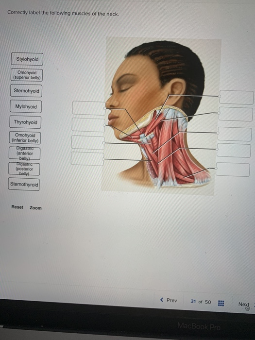
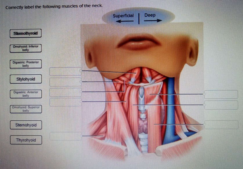
Correctly label the following muscles of the neck. Correctly label the following muscles of the neck SuperficialDeep Slemothyrod Omohyoid bely Digastnc. Stylohyoid Omohyoid superior belly Sternohyoid Mylohyoid Thyrohyoid inferior anterior belly posterior Sternothyroid Reset Zoom くPrev 31of 50Hİ Next.
Muscles coursing within the boundaries of the posterior neck triangle include the anterior middle and posterior scalene muscles as well as the omohyoid muscle. The suboccipital muscles act to rotate the head and extend the neck. It tilts the head to its own side and rotates the head so the head faces the opposite side.
The muscles that comprise the boundary of the posterior neck triangle in the sternocleidomastoid and trapezius muscles. Can you name the main anatomical areas of the brain. It attaches to the clavicle and scapula.
These muscles have two origins one on the sternum and the other on the clavicle. When both the splenius muscles act together they are what extend the head bring it head back. Superficial lymphatic drainage of the head and neck lateral view.
Humans have well-developed muscles in the face that permit a large variety of facial expressions. Try our top 10 quizzes. Tensor fasciae lata TFL originates on the anterior portion of the iliac crest and ASIS and inserts into the ITB.
Located underneath the platysma on the sides of the neck are the sternocleidomastoid muscles. There are also muscles that act on the hyoid and laryngeal skeleton sternohyoid sternothyroid thyrohyoid omohyoid stylohyoid and. How about the bones of the axial skeleton.
Do you know the bones of the skull. Muscles of the Neck and Head Labeling Printout. These are found in the back of the neck.
The platysma muscle is found overlying the triangle superficially. Several aspects of chewing swallowing and vocalizing are aided by eight pairs of hyoid muscles associated with. The trapezius is the most superficial muscle of the back and forms a broad flat triangle.
Correctly label the bones and anatomical features of the fetal. Anatomy and Physiology questions and answers. The skull reaches about three-quarters its adult size by age 1.
The muscles of the anterior region in front of the vertebral bodies and the muscles of. Correctly label the following muscles of the neck. With one on each side of the neck these help flex the neck and rotate the head upward and side to side.
The anterior neck muscles are a group of muscles covering the anterior aspect of the neck. The neck is divided into 4 regions to which some sub-regions or triangles belong. The lateral neck muscles also called the lateral vertebral muscles are a group of.
This division is based on the usually visible and or palpable borders of the large and relatively superficial sternocleidomastoid and trapezius muscles which are contained within the outermost investing layer of deep cervical fascia. One on each side of the neck. Name the parts of the human heart.
5 - the axial skeleton. Similarly what muscles are involved in neck extension. The four regions and their sub-triangles or.

Solved Correctly Label The Following Muscles Of The Neck Chegg Com

Neck Muscle Anatomy Health Medicine And Anatomy Reference Pictures Anatomiya Cheloveka Anatomiya Myshechnaya Sistema

11 4 Identify The Skeletal Muscles And Give Their Origins Insertions Actions And Innervations Anatomy Physiology

No comments for "Correctly Label the Following Muscles of the Neck."
Post a Comment